Facing a skin cancer diagnosis can be daunting. Whether your cancer requires excision, Mohs surgery coordination, or reconstructive repair, Dr. Gupta’s expertise will help you heal, while restoring both form and function.
Skin Cancer and Reconstruction San Diego

Patient Reviews
I’m amazed and grateful to say that what could have been a serious disfigurement is now, four months later, hardly noticeable
“I had a fairly large area of skin cancer removed from the side of my nose—about the size of a quarter. Dr. Gupta did a great job putting everything back together. I’m amazed and grateful to say that what could have been a serious disfigurement is now, four months later, hardly noticeable. Dr. Gupta is extremely skilled, and also personable and professional. Thank you, Dr. Gupta!”
5 star rating

Dr. Gupta performed the reconstruction, and I not only have no cancer, but I have my smile back!
“Dr. Gupta and his team are amazing. I had a serious squamous cell cancer treated with Mohs surgery, and I lost over half of my upper lip. Dr. Gupta performed the reconstruction, and I not only have no cancer, but I have my smile back!”
5 star rating

I was referred to Dr. Gupta by my dermatologist to repair my nose following Mohs surgery for skin cancer. Dr. Gupta rebuilt my nose, and I’m very pleased and relieved with the result
“Dr. Gupta rebuilt my nose, and I’m very pleased and relieved with the result. He was kind, highly capable, and instilled confidence throughout the entire process. From the initial consultation through surgery and post-op checkups, he was always prompt, courteous, and—most importantly—highly skilled and professional. I would highly recommend him.”
5 star rating

People were making comments like: 'You can’t even tell,' and 'He did an amazing job.'
“Dr. Gupta removed cancerous basal cells on my face; the procedure required ten stitches, and after two weeks, people were making comments like: 'You can’t even tell,' and 'He did an amazing job.”
5 star rating

I am very happy with the results!
“I had to patch up a hole in my head left from cancer surgery, and I am very happy with the results!”
5 star rating

Why Choose Dr. Gupta?
Board-Certified Plastic Surgeon for 25+ Years
Dr. Gupta has spent two decades refining his approach to reconstruction procedures and is extensively trained in microvascular techniques, flaps, grafts, and advanced repairs.
Personalized Care
From planning your procedure details to all your follow-up visits, Dr. Gupta is directly involved with your care every step of the way.
Natural Artistry
You can trust Dr. Gupta to approach your reconstruction in a way that preserves function and enhances form.

The Benefits of Skin Cancer Reconstruction
- Restore tissue in areas where skin was removed during cancer treatment
- Preserve facial symmetry, contour, and function after skin cancer removal
- Provide emotional closure and improved confidence after your diagnosis and treatment

The Skin Cancer and Reconstruction Process
When you’re diagnosed with skin cancer, your first step is to schedule consultations with your dermatologist, Mohs surgeon, and plastic surgeon. Dr. Gupta works closely with your other providers during the planning stages to evaluate the defect, surgical margins and available local tissue. Using diagrams, he’ll help you understand your options, including grafts, local flaps, and complex flaps, and how each technique will impact your appearance and recovery.
In many cases, Dr. Gupta will perform the reconstruction immediately after cancer removal. He can harvest tissue from nearby areas and use flaps or grafts to expertly close the defect, reconnect a blood supply, and contour tissue to match the surrounding skin. Throughout this process, he places incisions strategically to ensure your resulting scars are hidden within your body’s natural folds.
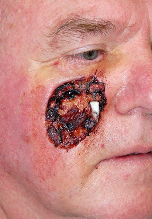
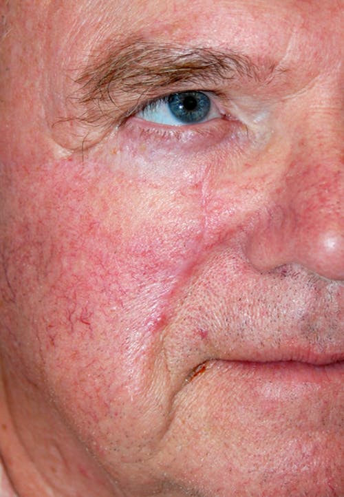
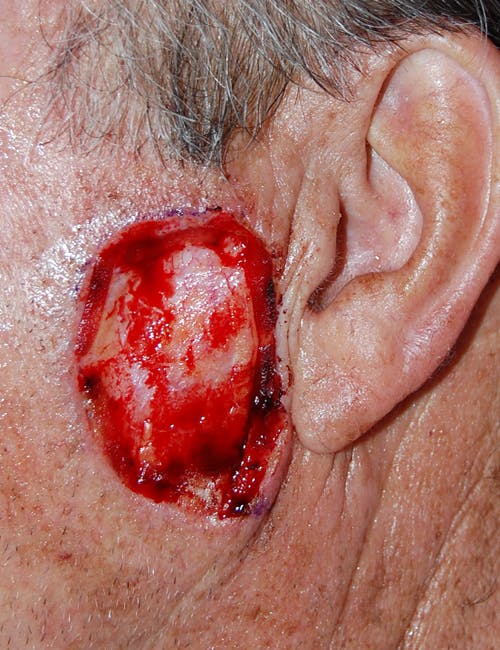
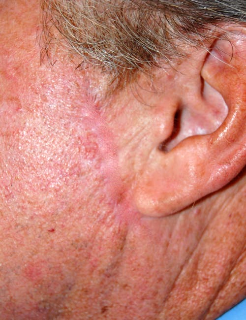
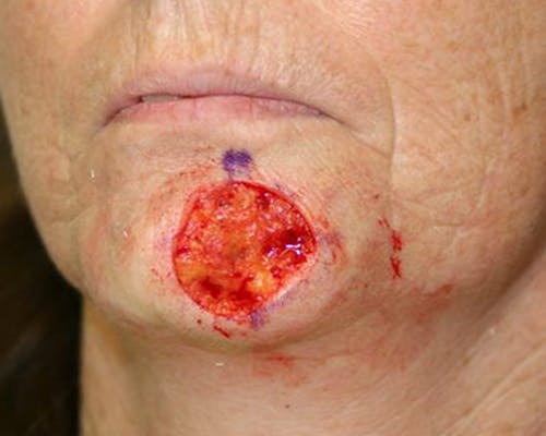
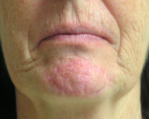
Mohs Chin Surgery Before & After
Patient Reviews
“Dr. Gupta is wonderful. He has such a calm bedside manner, and explains everything in easy to understand detail. He is doing a great job reconstructing my breasts after a devastating loss to cancer, and I always feel better about myself after an appointment. His staff makes you feel welcome from the minute you open the door. He is simply the best.”
5 star rating
“I am grateful to Dr. Gupta and his staff for the positive experience and outcome of my recent procedure. Dr. Gupta's kind confidence set me at ease from the first appointment. Sarah and Reagan are amazing in the office. So responsive and positive! I highly recommend Dr. Gupta!”
5 star rating
“I just received a birthday card from Dr. Gupta. I was his patient seventeen years ago, for breast reconstruction, after having breast cancer. Today, when I visit doctors for exams, they tell me, 'It’s the best breast reconstruction I’ve ever seen!'”
5 star rating
“I had a breast mastopexy done and I couldn’t be any happier with my results. Dr. Gupta and Sarah are a great team and are super easy to get ahold of, very helpful. I feel so relieved that my back pain will be gone and I will be able to feel more confident in my body! Huge thanks to Dr. Gupta.”
5 star rating
“Dr. Gupta is wonderful and takes his time with his patient and explains things thoroughly. Not rushed. Front desk is always welcoming and thoughtful.”
5 star rating
“Dr. Gupta was personable, knowledgeable and helpful. He took time to explain my injury and the best course of action going forward.”
5 star rating
“Best experience ever. I got a breast reduction and I’m so happy with the results. Dr. Gupta is great and the office staff are great.”
5 star rating
“Dr. Gupta and staff were accommodating and responsive to my needs. Surgery was successful with no issues. Very positive experience. Highly recommended!”
5 star rating
“From the receptionist to the doctor everyone was very polite and knowledgeable. I was very happy with my experience. Dr. Gupta explained everything and answered all my questions. I am excited to move forward with my surgery. I would recommend him to anyone!”
5 star rating
“As with every visit, Dr. Gupta is such an easy surgeon to work with. He asks pertinent questions related to me and my recovery. Each question I ask us answered with explanations and details. Sara and Courtney are both friendly and helpful. I have recommended Dr Gupta to numerous friends. On a scale of 10, I would give Dr. Gupta a 15/10. I feel like he truly cares about me and my recovery.”
5 star rating
“He is very kind and informative, and made me feel comfortable. He answered all of my questions thoroughly and with drawings or demonstrations. Dr. Gupta addressed my concerns and I left feeling secure about my decision to move forward with my surgery. His office is beautiful and his staff was professional and informative regarding pricing and financing options.”
5 star rating
“Dr. Gupta is an Exceptional Dr. with incredible attention to detail and a consummate Perfectionist! He is professional, personable and extremely experienced! I trust him implicitly. His staff is kind and professional and very helpful. I sincerely cannot recommend him more highly!”
5 star rating
“Dr. Gupta is wonderful. He has such a calm bedside manner, and explains everything in easy to understand detail. He is doing a great job reconstructing my breasts after a devastating loss to cancer, and I always feel better about myself after an appointment. His staff makes you feel welcome from the minute you open the door. He is simply the best.”
5 star rating
“His surgical skill and attention to detail are exceptional.”
““As a board-certified dermatologist, I frequently refer patients to Dr. Gupta for reconstructive procedures following Mohs surgery and for excision of melanoma with sentinel lymph node biopsy. His surgical skill and attention to detail are exceptional, and the quality of his results consistently exceeds expectations. Most of my patients specifically request Dr. Gupta when they require treatm...”
“As a board-certified dermatologist, I frequently refer patients to Dr. Gupta for reconstructive procedures following Mohs surgery and for excision of melanoma with sentinel lymph node biopsy. His surgical skill and attention to detail are exceptional, and the quality of his results consistently exceeds expectations. Most of my patients specifically request Dr. Gupta when they require treatment for new skin cancers, reflecting the trust and confidence he inspires.”
5 star rating
“He is a master surgeon.”
““I have had the pleasure of working with Dr. Abhay Gupta for many years. He is a very special surgeon and a very special person. He is a master surgeon. I send my patients and my family to him whenever needed. I highly recommend him.””
5 star rating
“A plastic surgeon of excellence.”
““I have worked with Dr. Gupta for many years and twice myself have been his patient for excision of cancerous skin lesions. As a practicing anesthesiologist, I have worked with hundreds of different surgeons over the past forty years, and Dr. Gupta is one of the best! I strongly recommend Dr. Gupta as a plastic surgeon of excellence.””
5 star rating
“His results are consistently beautiful.”
““I’ve known and worked with Dr. Gupta for more than 15 years, and I’ve seen firsthand the care and attention he gives his patients. His results are consistently beautiful, and he also has a kind and genuine bedside manner. I trust him completely and even sent my own family members to him. Happy to have him as a colleague.””
5 star rating
“His results speak for themselves.”
““Dr. Gupta is an exceptional plastic surgeon and a true gem in our medical community. His professionalism, compassion, and meticulous care set the standard—I trust him implicitly and have referred both patients and family to him with complete confidence. He consistently goes above and beyond, and his results speak for themselves.””
5 star rating
“Dr Gupta exudes professionalism, and he demonstrates great care for both his cosmetic and non-cosmetic patients.”
5 star rating
“We know our patients will be well taken care of when referred to Gupta Plastic Surgery.”
5 star rating
He has always exhibited the utmost quality for care...
“I have known Doctor Gupta for over fifteen years and he has always exhibited the utmost quality for care for his patients. Professionalism, Empathy, and Kindness are core tenets of his practice, and I am honored to call him one of my colleagues.”
5 star rating
“I’ve worked closely with Dr. Gupta and can speak to his surgical skill and strong work ethic.
He is a trusted colleague and a true professional.”
5 star rating
An exceptionally skilled and dedicated plastic surgeon...
“Dr. Abhay Gupta is an exceptionally skilled and dedicated plastic surgeon whose meticulous technique and compassionate approach consistently earn the admiration of both colleagues and patients. His commitment to excellence and innovation in the field sets a high standard, and it's a privilege to work alongside someone of his caliber.”
5 star rating
Our patients come back raving about his skill...
“We are so lucky to have Dr. Gupta in our community. We always have so much confidence in his repairs after Mohs surgery. Our patients come back raving about his skill and bedside manner, and that means everything!”
5 star rating
“Dr Gupta is a superb plastic surgeon with whom I have had the pleasure of collaborating with for over 20 years. Reconstructive and cosmetic patients are extremely happy with his care and outstanding work!”
5 star rating
“Dr. Gupta was the 2nd plastic surgeon that I consulted for my tummy tuck and breast lift/augmentation. I lost over 100 lbs., the old fashioned way and the 1st surgeon I met with treated me awful! He found out I was a long time smoker and saw the amount of skin from the weight loss that needed to be dealt with, and he just lost it! That Dr. made me feel horrible about my body. I finally went to see...”
Dr. Gupta was the 2nd plastic surgeon that I consulted for my tummy tuck and breast lift/augmentation. I lost over 100 lbs., the old fashioned way and the 1st surgeon I met with treated me awful! He found out I was a long time smoker and saw the amount of skin from the weight loss that needed to be dealt with, and he just lost it! That Dr. made me feel horrible about my body. I finally went to see Dr. Gupta and was terrified I would get the same reaction. I did NOT!! Dr. Gupta was so wonderful and professional. He worked with me to help me quit smoking and assured me that we would have great results. I am 40 yrs old and a mother of 3 and I look fabulous!! Dr. Gupta is amazing! I need to give a huge compliment to his staff and especially the Encinitas Surgery Center! I was treated like a princess at every visit.
5 star rating
“Went to three other surgeons before finding Dr. Gupta, and this is the most profesional office I have found. I am so excited, I had financial issues, and the office worked with me to find the necessary financing. Paulette and the staff are wonderful. Upon checking the website, I found that Dr. Gupta’s credentials were outstanding in surgery–both general and cosmetic. I am scheduled in ...”
Went to three other surgeons before finding Dr. Gupta, and this is the most profesional office I have found. I am so excited, I had financial issues, and the office worked with me to find the necessary financing. Paulette and the staff are wonderful. Upon checking the website, I found that Dr. Gupta’s credentials were outstanding in surgery–both general and cosmetic. I am scheduled in January.
5 star rating
“I had basal cell skin cancer on the end of my nose. I feel so blessed to meet a Doctor with such patience and honesty. He sent me to a great MOHs surgeon who was patient and took his time. The next morning I was scheduled for Dr. Gupta and reconstructive surgery. At 6:30 am I was dressed and ready to start with my wife by my side. Dr Gupta walks in to examine the wound and consult me on the proced...”
I had basal cell skin cancer on the end of my nose. I feel so blessed to meet a Doctor with such patience and honesty. He sent me to a great MOHs surgeon who was patient and took his time. The next morning I was scheduled for Dr. Gupta and reconstructive surgery. At 6:30 am I was dressed and ready to start with my wife by my side. Dr Gupta walks in to examine the wound and consult me on the procedure. He calmly examines my wound from the MOHs surgery. To our surprise he says I have two options for you. Have the surgery or follow my wound care instructions and your nose will heal by itself. I asked him what would he do? ” He said if I were you I would go home and start wearing that Band-aid and bacitracin 24 hours a day for the next month.” Thanks to Dr. Gupta’s honesty my nose is almost healed three months later without the plastic surgery. He has truly exceeded my expectations as a Doctor and a person I truly trust.
5 star rating
“Dr Gupta and his staff are the most professional and yet friendly organization that I have ever associated with. You never have to wait in the office, you are seen by Dr Gupta immediately, you are given a thorough explanation of what you are asking to have done and then you walk out knowing the total amount you will be required to spend. Everything is very nicely operated. This will be my second s...”
Dr Gupta and his staff are the most professional and yet friendly organization that I have ever associated with. You never have to wait in the office, you are seen by Dr Gupta immediately, you are given a thorough explanation of what you are asking to have done and then you walk out knowing the total amount you will be required to spend. Everything is very nicely operated. This will be my second surgery with Dr Gupta and I couldn’t be in better hands.
5 star rating
“I would highly recommend Dr. Gupta to a friend or colleague. I had breast cancer last year with a double mastectomy. When I saw Dr. Gupta a couple of months after my mastectomy, he was very professional and recommended that I wait until I am further healed and feel more comfortable for reconstruction surgery to be done. I sincerely respect Dr. Gupta for his honesty and caring for his patients. Dr....”
I would highly recommend Dr. Gupta to a friend or colleague. I had breast cancer last year with a double mastectomy. When I saw Dr. Gupta a couple of months after my mastectomy, he was very professional and recommended that I wait until I am further healed and feel more comfortable for reconstruction surgery to be done. I sincerely respect Dr. Gupta for his honesty and caring for his patients. Dr. Gupta is honest, sincere, precise.
5 star rating
“Dr. Gupta is an extremely talented plastic surgeon. My results have been beyond my expectation. I am very happy and look forward to seeing him & his great staff again in the near future!”
5 star rating
“I’ve had large breasts since i was 11 years old .. Which obviously was creating some major back problems, and huge discomfort! It was always really hard to exercise and work out- due to the pain i would get – from all the weight on my shoulders… My primary Dr. referred me to Dr. Gupta for a breast reduction consultation. I had planned to consult with several plastic surgeons to ...”
I’ve had large breasts since i was 11 years old .. Which obviously was creating some major back problems, and huge discomfort! It was always really hard to exercise and work out- due to the pain i would get – from all the weight on my shoulders… My primary Dr. referred me to Dr. Gupta for a breast reduction consultation. I had planned to consult with several plastic surgeons to see my options. But as soon as i met with Dr. Gupta, I knew i didn’t have to waste my time to see someone else! Aside from the very impressive photos/portfolio on his website he put my mind at ease. I was barely 20 years old when i had my surgery done, so you can imagine how nervous and worried i was. But i have to say, it was the best decision of my life! I am so happy with the results, and feel amazing! I was very satisfied with the care and catering from himself and his office staff. I will refer everyone to Dr. Gupta!
5 star rating
“Dr. Gupta is one of the best plastic surgeons in San Diego. I had bad breast surgery by another surgeon who refused to listen to any of my complaints. I was referred to Dr. Gupta by a friend and was very impressed immediately. He put me at ease right away and answered all my questions. Excellent bedside manner. He fixed my breasts with one operation and now they look great. I would highly recommen...”
Dr. Gupta is one of the best plastic surgeons in San Diego. I had bad breast surgery by another surgeon who refused to listen to any of my complaints. I was referred to Dr. Gupta by a friend and was very impressed immediately. He put me at ease right away and answered all my questions. Excellent bedside manner. He fixed my breasts with one operation and now they look great. I would highly recommend him to anyone.
5 star rating
“Dr. Gupta is everything his reputation says he is. He has the BEST bedside manner of any doctor I have ever seen. He explains EVERYTHING to you about the surgery and answers EVERY question. He is so calm and relaxing from the very first consultation to every follow up appointment. His staff is wonderful. The office is beautiful, I love the waterfall in the reception lobby. My surgery went perfect ...”
Dr. Gupta is everything his reputation says he is. He has the BEST bedside manner of any doctor I have ever seen. He explains EVERYTHING to you about the surgery and answers EVERY question. He is so calm and relaxing from the very first consultation to every follow up appointment. His staff is wonderful. The office is beautiful, I love the waterfall in the reception lobby. My surgery went perfect and I healed great. I can barely even see the scars. Thanks Dr. Gupta, I’m definitely coming back to you for my next surgery.
5 star rating
“Dr Gupta is amazing! He did my breast implants and tummy tuck. He took the time to explain everything to me. And then the operation was a huge success! I’ll never go to anyone else. Dr Gupta is the best.”
5 star rating
“Dr. Gupta was the 2nd plastic surgeon that I consulted for my tummy tuck and breast lift/augmentation. I lost over 100 lbs., the old fashioned way and the 1st surgeon I met with treated me awful! He found out I was a long time smoker and saw the amount of skin from the weight loss that needed to be dealt with, and he just lost it! That Dr. made me feel horrible about my body. I finally went to see...”
Dr. Gupta was the 2nd plastic surgeon that I consulted for my tummy tuck and breast lift/augmentation. I lost over 100 lbs., the old fashioned way and the 1st surgeon I met with treated me awful! He found out I was a long time smoker and saw the amount of skin from the weight loss that needed to be dealt with, and he just lost it! That Dr. made me feel horrible about my body. I finally went to see Dr. Gupta and was terrified I would get the same reaction. I did NOT!! Dr. Gupta was so wonderful and professional. He worked with me to help me quit smoking and assured me that we would have great results. I am 40 yrs old and a mother of 3 and I look fabulous!! Dr. Gupta is amazing! I need to give a huge compliment to his staff and especially the Encinitas Surgery Center! I was treated like a princess at every visit.
5 star rating
“Went to three other surgeons before finding Dr. Gupta, and this is the most profesional office I have found. I am so excited, I had financial issues, and the office worked with me to find the necessary financing. Paulette and the staff are wonderful. Upon checking the website, I found that Dr. Gupta’s credentials were outstanding in surgery–both general and cosmetic. I am scheduled in ...”
Went to three other surgeons before finding Dr. Gupta, and this is the most profesional office I have found. I am so excited, I had financial issues, and the office worked with me to find the necessary financing. Paulette and the staff are wonderful. Upon checking the website, I found that Dr. Gupta’s credentials were outstanding in surgery–both general and cosmetic. I am scheduled in January.
5 star rating
“I absolutely love the results of my breast augmentation and liposuction. I was referred to Dr. Gupta’s ex-partner by a friend and consulted with him prior to Dr. Gupta. I wasn’t impressed at all with the other doctor. I felt like I was being rushed between patients and he didn’t spend enough time discussing the procedure. Not to mention his scheduler, she didn’t have a warm...”
I absolutely love the results of my breast augmentation and liposuction. I was referred to Dr. Gupta’s ex-partner by a friend and consulted with him prior to Dr. Gupta. I wasn’t impressed at all with the other doctor. I felt like I was being rushed between patients and he didn’t spend enough time discussing the procedure. Not to mention his scheduler, she didn’t have a warm personality at all. I was amazed by the difference in Dr. Gupta’s bed-side manner, professionalism and honesty. I know I would have regretted having surgery with the other doctor , especially after reading his reviews on this same site. I’m extremely happy with my decision to have surgery with Dr. Gupta and would recommend him to any one.
5 star rating
“Dr. Gupta is amazing – professional, caring, skilled considerate. Answers ALL of my questions and I never felt
rushed with them. Every physician in San Diego should take lessons from him.”
5 star rating
“Dr. Gupta and the team he has behind him are the best! I feel fortunate to be a patient of Dr. Gupta. Due to breast cancer I have been going to Dr. Gupta since this past December. He and his staff help to make you comfortable and relaxed during a stressfull period. He is easy to get a hold of if necessary. Appointments pass quickly with little time in the waiting room. Dr. Gupta is excellent and a...”
Dr. Gupta and the team he has behind him are the best! I feel fortunate to be a patient of Dr. Gupta. Due to breast cancer I have been going to Dr. Gupta since this past December. He and his staff help to make you comfortable and relaxed during a stressfull period. He is easy to get a hold of if necessary. Appointments pass quickly with little time in the waiting room. Dr. Gupta is excellent and always appears to look forward to seeing you as you look forward to seeing him and his nice smile.
5 star rating
“Dr. Gupta is a wonderful doctor. He is compassionate, caring and extremely skilled. I have nothing but praise for Dr. Gupta. He has always been accessible and responsive to my needs. He has provided excellent care during my post breast cancer reconstruction. I highly recommend him as a surgeon.”
5 star rating
“Dr. Gupta is the best. He did my breast implants and liposuction a few months ago and I love my results. He always had a smile on his face and he always had the time to answer my questions. It was very comforting to know that if I ever needed to talk to him, he was always available on his cell phone, even at night. His office staff are really nice. I’m glad I found such a great plastic surge...”
Dr. Gupta is the best. He did my breast implants and liposuction a few months ago and I love my results. He always had a smile on his face and he always had the time to answer my questions. It was very comforting to know that if I ever needed to talk to him, he was always available on his cell phone, even at night. His office staff are really nice. I’m glad I found such a great plastic surgeon, and I’m not going to go anywhere else.
5 star rating
“Dr. Gupta did my breast reconstruction after my double mastectomy. He is a delightful person, with a professional manner which instills confidence. He provides a lengthy print-out explaining what would be involved and what to expect, etc., and is totally open to answering any and all questions. His staff, by the way, is super, knowledgeable, and helpful. AND, the results are fantastic. I couldn&rs...”
Dr. Gupta did my breast reconstruction after my double mastectomy. He is a delightful person, with a professional manner which instills confidence. He provides a lengthy print-out explaining what would be involved and what to expect, etc., and is totally open to answering any and all questions. His staff, by the way, is super, knowledgeable, and helpful. AND, the results are fantastic. I couldn’t be more pleased with the reconstruction. I highly recommend Dr. Abhay Gupta.
5 star rating
“I was quite scared about getting breast reconstruction done, since having a mastectomy 11 yrs before. Dr. Gupta made me feel at ease and he was extremely caring and compassionate also. My husband and I had many questions to ask and Dr. Gupta answered them all in a way we could understand, and most importantly, he did not seem to be in a hurry. He was always pleasant to talk to and even when I had ...”
I was quite scared about getting breast reconstruction done, since having a mastectomy 11 yrs before. Dr. Gupta made me feel at ease and he was extremely caring and compassionate also. My husband and I had many questions to ask and Dr. Gupta answered them all in a way we could understand, and most importantly, he did not seem to be in a hurry. He was always pleasant to talk to and even when I had the injections to fill the expander, he would reassure me each time that all was going well. At nearly 60, this was very important to me. For the first part of the reconstruction surgery, Dr. Gupta actually wheeled me into the surgery area… what other doctor would do that?? Dr. Gupta’s staff was also the greatest, working around my busy schedule with the foster kids. I would refer anyone to Dr. Gupta. He is the BEST!
5 star rating
“I was referred to Dr. Gupta for an evaluation for abdominal reconstructive surgery and cosmetic repair as a result of multiple surgeries resulting from a ruptured appendix, subsequent surgical procedures, and multiple hernias. I could not be happier with the result. Dr. Gupta is the consummate medical professional, obtaining the very best result from these procedures. He and his office staff alway...”
I was referred to Dr. Gupta for an evaluation for abdominal reconstructive surgery and cosmetic repair as a result of multiple surgeries resulting from a ruptured appendix, subsequent surgical procedures, and multiple hernias. I could not be happier with the result. Dr. Gupta is the consummate medical professional, obtaining the very best result from these procedures. He and his office staff always respected my time, and I was seen at the appointed time. I am not planning on any future procedures with Dr. Gupta, but recognize that future needs may occur, and I would call upon Dr. Gupta’ professional services as needed. I have the utmost faith in his expertise, and would offer any level of recommendation to others seeking his level of expertise.
5 star rating
“Dr. Gupta is the third plastic surgeon I have used in the San Diego area and I will NEVER look for another. He is caring, skilled, patient… He completed reconstructive breast surgery begun by another doctor earlier this year, as well as some abdominal work. He made really great recommendations for a small amount of additional work he felt would be beneficial, then gave me time to decide. Pr...”
Dr. Gupta is the third plastic surgeon I have used in the San Diego area and I will NEVER look for another. He is caring, skilled, patient… He completed reconstructive breast surgery begun by another doctor earlier this year, as well as some abdominal work. He made really great recommendations for a small amount of additional work he felt would be beneficial, then gave me time to decide. Pre-op explanations were clear and relevant and my entire experience was positive from first visit to all post-ops had to date. My post-surgical pain was well maintained. My results are better than I could have imagined. His office staff is also great to work with. Unfortunately, I have been one of those patients who remembers “one more question” as soon as my appointment is over. They have been unfailingly pleasant and gotten me an answer within moments. Dr. Gupta himself has never hesitated to return to the office or the phone to answer my questions. DEFINITELY RECOMMEND DR GUPTA AND HIS STAFF.
5 star rating
“After the damage that was done by having four children, including a set of twins, I finally decided to have an abdominoplasty in order to return to my pre-pregnancy condition. I could not be happier with the results. It has been a year, and I feel wonderful and even now, when I run into people I haven’t seen in a while, they are shocked and amazed. Thank You Dr. Gupta!”
5 star rating
“Dr. Gupta is extremely knowledgeable on the subject of plastic surgery. I saw him for repairs due to breast cancer treatment. He was kind, well versed and answered all of my questions. In many cases he answered the question before I even asked. He was understanding, patient and made my decision easy. He was honest about all the possible outcomes of my surgery, quickly returned calls during my reco...”
Dr. Gupta is extremely knowledgeable on the subject of plastic surgery. I saw him for repairs due to breast cancer treatment. He was kind, well versed and answered all of my questions. In many cases he answered the question before I even asked. He was understanding, patient and made my decision easy. He was honest about all the possible outcomes of my surgery, quickly returned calls during my recovery time as well. His staff is very helpful too. I would refer close friends and family to him!
5 star rating
“Dr. Gupta is an excellent doctor, I would recommend him to anyone and everyone! I am very happy with the results of my reconstructive surgery. During a very difficult time in my life, Dr. Gupta was always kind, compassionate, patient, and knowledgeable! He always took time to address any questions and/or concerns that I had. Thank you Dr. Gupta!”
5 star rating
“LOVE Dr. Gupta and his staff! I will always be a loyal patient to Dr. Gupta! I have been to a big plastic surgery office in La Jolla and I didn’t feel any one on one attention while I was there. After going to Dr. Gupta’s my experience and my opinion of Plastic surgery offices has completely changed for the better! No other Doc has made me feel so comfortable and welcome. He is extreme...”
LOVE Dr. Gupta and his staff! I will always be a loyal patient to Dr. Gupta! I have been to a big plastic surgery office in La Jolla and I didn’t feel any one on one attention while I was there. After going to Dr. Gupta’s my experience and my opinion of Plastic surgery offices has completely changed for the better! No other Doc has made me feel so comfortable and welcome. He is extremely professional and the office is so relaxing and beautiful!
5 star rating
“Dr. Gupta performed a nose reconstruction after a MOHS surgery to remove a Basel cell. He was quite pleasant and professional at my first visit and the procedure. He explained the steps thoroughly and prepared me for what to expect pre and post surgery. His assistant, Paulette, was very helpful in getting all the information lined up from copies of bloodwork, other doctor reports and releases. Suc...”
Dr. Gupta performed a nose reconstruction after a MOHS surgery to remove a Basel cell. He was quite pleasant and professional at my first visit and the procedure. He explained the steps thoroughly and prepared me for what to expect pre and post surgery. His assistant, Paulette, was very helpful in getting all the information lined up from copies of bloodwork, other doctor reports and releases. Such steps that I was not prepared to persue due to my lack of experience and age. I would highly recommend Dr. Gupta and his team.
5 star rating
“The entire process with Dr Gupta and his staff has been excellent. Surgery, follow up and office visits all highly professional. Results are better than expected. Highly recommend this practice.”
5 star rating
“I appreciate everything that Dr. Gupta and his staff have done for me. Paulette, Kamryn and Dr. Gupta each demonstrated the highest level of professionalism. My procedure went smoothly. I anticipate that my recovery will too. Best regards to all of you.”
5 star rating
“My appointment was right on time. My questions were answered. I feel heard and well taken care of. Thank you!”
5 star rating
“Dr Abhay is the best! As a guy, I was embarrassed to seek help on what I was self conscious about. I was immediately put at easy by his team and him. He listen with no judgment and was interested not only my concerns, but ME! I highly recommend Dr. Gupta!”
5 star rating
“Dr. Gupta was personable, knowledgeable and helpful. He took time to explain my injury & the best course of action going forward.”
5 star rating
The outcome of what Dr. Gupta did is phenomenal
“It has been four weeks since my surgery, and I feel amazing. I have been cleared to start working out, and the outcome of what Dr. Gupta did is phenomenal.”
5 star rating
It was the best surgery I have ever had
“Dr. Gupta did my tummy tuck and breast lift, and it was the best surgery I have ever had. If you want to get plastic surgery, go see Dr. Gupta – he’s the best.”
5 star rating
I am 40 years old and a mother of 3, and I look fabulous!!
“Dr. Gupta was the 2nd plastic surgeon that I consulted for my tummy tuck and breast lift/augmentation... I am 40 years old and a mother of 3, and I look fabulous!!”
5 star rating
I could not be happier with the result
“I was referred to Dr. Gupta for an evaluation for abdominal reconstructive surgery and cosmetic repair as a result of multiple surgeries... I could not be happier with the result.”
5 star rating
I was thrilled
“When I realized... Dr. Gupta and staff [were willing] to accommodate a separate financial plan for me to combine cosmetic surgery (tummy tuck) with my need for multiple hernia repairs... I was thrilled.”
5 star rating
I can't believe I can finally wear a bikini again after having my two kids
“Dr. Gupta is an amazing surgeon! I had a breast augmentation and tummy tuck 3 months ago, and I am ecstatic with my new body. Dr. Gupta really took the time to answer all my questions and made me feel so comfortable during my appointments. He is extremely caring and I am very grateful for everything he has done for me. I can't believe I can finally wear a bikini again after...”
Dr. Gupta is an amazing surgeon! I had a breast augmentation and tummy tuck 3 months ago, and I am ecstatic with my new body. Dr. Gupta really took the time to answer all my questions and made me feel so comfortable during my appointments. He is extremely caring and I am very grateful for everything he has done for me. I can't believe I can finally wear a bikini again after having my two kids. Thanks Dr. Gupta!!
5 star rating
Dr. Gupta is everything his reputation says he is
“Dr. Gupta is everything his reputation says he is. He has the BEST bedside manner of any doctor I have ever seen. He explains EVERYTHING to you about the surgery and answers EVERY question. He is so calm and relaxing from the very first consultation to every follow-up appointment. His staff is wonderful. My surgery went perfectly, and I healed great. I can barely even see the scars. Thanks, Dr. Gu...”
Dr. Gupta is everything his reputation says he is. He has the BEST bedside manner of any doctor I have ever seen. He explains EVERYTHING to you about the surgery and answers EVERY question. He is so calm and relaxing from the very first consultation to every follow-up appointment. His staff is wonderful. My surgery went perfectly, and I healed great. I can barely even see the scars. Thanks, Dr. Gupta, I’m definitely coming back to you for my next surgery.
5 star rating
Dr. Gupta is amazing! He did my breast implants and tummy tuck
“Dr. Gupta is amazing! He did my breast implants and tummy tuck. He took the time to explain everything to me. I’ll never go to anyone else.”
5 star rating
Super satisfied with my results
“I had a tummy tuck and breast implants done by Dr. Gupta and am super satisfied with my results. I’m very thankful, and I am a patient for life.”
5 star rating
Dr. Gupta made me feel really relaxed with his relaxed demeanor
“I was referred to him by a friend when I was looking to get a tummy tuck and breast lift. Dr. Gupta made me feel really relaxed with his relaxed demeanor and incredible bedside manner.”
5 star rating
I have to say that Dr. Gupta is the best that I have ever seen in my career
“I am a nurse at a hospital in San Diego and I also work at a busy surgery center. Over the past few years, I have worked with about 25 different plastic surgeons in the operating room. I have to say that Dr. Gupta is the best that I have ever seen in my career. His results are amazing, and his hands are truly gifted.”
5 star rating
It’s only been 5 days since my surgery, and WOW, the results
“It’s only been 5 days since my surgery, and WOW, the results. Dr. Gupta is excellent. Even the girls who assisted him made me very comfortable and relaxed. I would recommend Dr. Gupta to all I know who would benefit from his expertise.”
5 star rating
I deeply researched and then met with multiple surgeons before meeting Dr. Gupta
“I deeply researched and then met with multiple surgeons before meeting Dr. Gupta and his staff. I was immediately impressed with how comfortable I felt. The environment was therapeutic, and Dr. Gupta's bedside manner was impeccable.”
5 star rating
Dr. Gupta is warm, professional, kind, and considerate
“Dr. Gupta is warm, professional, kind, and considerate. Plastic surgery is not just about any old doctor... It can also be a skill that very few possess that is required to literally save your life and reconstruct the broken or torn pieces. Dr. Gupta takes the time to learn far beyond the easy stuff and develops skill sets that can not only change a life but save it.”
5 star rating
Every physician in San Diego should take lessons from him
“Dr. Gupta is amazing – professional, caring, skilled, and considerate. He answers ALL of my questions, and I never felt rushed with them. Every physician in San Diego should take lessons from him.”
5 star rating
I went to three other surgeons before finding Dr. Gupta
“I ent to three other surgeons before finding Dr. Gupta, and this is the most professional office I have found. Upon checking the website, I found that Dr. Gupta’s credentials were outstanding in surgery, both general and cosmetic.”
5 star rating
I couldn't have asked for a better experience and result
“I couldn't have asked for a better experience and result. Dr. Gupta is professional, caring and definitely puts the best interest of his patients first! I highly recommend him for your plastic surgery and/or reconstructive needs.”
5 star rating
Thorough, informational, professional
“Thorough, informational, professional. I appreciated the staff and Dr. Gupta.”
5 star rating
I highly recommend Dr Gupta to anyone looking for a great doctor
“Dr Gupta and his staff are always patient and answer all the questions you may have. I highly recommend Dr Gupta to anyone looking for a great doctor.”
5 star rating
Dr. Gupta and his staff are great
“Dr. Gupta and his staff are great. I am always impressed with the professionalism and calming atmosphere of the office, and of course, the results! Thanks, Dr. Gupta!”
5 star rating
Amazing and excellent surgeon
“Amazing and excellent surgeon. I am so happy. Choose him!”
5 star rating
Dr. Gupta is an extremely talented plastic surgeon
“Dr. Gupta is an extremely talented plastic surgeon. My results have been beyond my expectations. I am very happy and look forward to seeing him & his great staff again in the near future!”
5 star rating
Best Plastic Surgeon you can find in San Diego!
“Best Plastic Surgeon you can find in San Diego! The Doctor is amazing and gives great results! Love the office and staff as well. If you want good quality work and service, then go to Gupta Plastic Surgery! Love Them!”
5 star rating
I felt safe and confident
“I felt safe and confident everything would go as planned.”
5 star rating
Dr. Gupta’s kind confidence set me at ease from the first appointment
“Dr. Gupta’s kind confidence set me at ease from the first appointment. Sarah is amazing in the office.”
5 star rating
I am and have been completely satisfied
“I am and have been completely satisfied and happy to have him do any procedures for me.”
5 star rating
I absolutely love the results of my breast augmentation and liposuction
“I absolutely love the results of my breast augmentation and liposuction... I’m extremely happy with my decision to have surgery with Dr. Gupta and would recommend him to anyone.”
5 star rating
I feel like a new person thanks to you
“You are an amazing surgeon and did an amazing job with my breast surgery. I am extremely happy with the results. I feel like a new person thanks to you.”
5 star rating
Dr. Gupta is the best
“Dr. Gupta is the best. He did my breast implants and liposuction a few months ago, and I love my results.”
5 star rating
Dr Gupta not only was supportive but made me feel very at ease
“Dr Gupta not only was supportive but made me feel very at ease with a bad situation... I've been told by other mastectomy victims what a great job he did on my surgery matching my built to my natural breast.”
5 star rating
Dr. Gupta is an expert in his field and my surgery went flawlessly
“Dr. Gupta is an expert in his field and my surgery went flawlessly. I had minimal pain and also have minimal scarring. Scheduling staff were very accommodating and friendly as well. I'm grateful to have had Dr. Gupta and staff to take care of me.”
5 star rating
I'm so glad I chose Dr. Gupta. I had the best experience! I would definitely recommend him!!
“I had a breast augmentation with very little breast tissue. I was worried they would look too unnatural but they healed amazingly and have a very natural look to them. Everything went smoothly with the surgery and the consultations. This was also my very first time having any kind of surgery, I'm so glad I chose Dr. Gupta. I had the best experience! I would definitely recommend him!!”
5 star rating
Dr. Gupta, Thank you so much for making a lifetime dream into reality for me
“Thank you so much for making a lifetime dream into reality for me. You are an amazing surgeon and did an amazing job with my breast surgery. I am extremely happy with the results. I feel like a new person thanks to you.”
5 star rating
Love the results of my new breast augmentation and lift!
“Very sweet Dr, always made me feel comfortable about my procedure and healing process. Love the results of my new breast augmentation and lift! I tell all my friends about him!”
5 star rating
I am very and extremely impressed with my results
“I had my breast augmentation done here almost 3 years ago. I am extremely impressed with my results and with how amazing the doctor was. He is extremely smart and knows what he is doing. I would absolutely recommend Dr. Gupta for any cosmetic surgery. I was very impressed with my results and, in the future, when I get another breast augmentation, I will absolutely go to the same cosmetic surgeon.”
I had my breast augmentation done here almost 3 years ago. I am extremely impressed with my results and with how amazing the doctor was. He is extremely smart and knows what he is doing. I would absolutely recommend Dr. Gupta for any cosmetic surgery. I was very impressed with my results and, in the future, when I get another breast augmentation, I will absolutely go to the same cosmetic surgeon.
5 star rating
I would HIGHLY recommend him!!!
“I am so pleased with his bedside manner, his staff, and the incredible job he did on my surgery. I would HIGHLY recommend him!!!”
5 star rating
I am so HAPPY with my result!
“Dr Gupta is a very talented surgeon. I recently had my surgery done by him, and I am so HAPPY with my result!”
5 star rating
I would not hesitate to recommend Dr. Gupta to every woman considering breast reconstruction, or any other surgery. He is absolutely fantastic and has changed my life!
“After researching and consulting with top plastic surgeons in both San Diego and Los Angeles, I found Dr. Gupta to be the most competent and knowledgeable. Dr. Gupta performed surgeries that not only eliminated my pain, but made my breasts look beautiful! I would not hesitate to recommend Dr. Gupta to every woman considering breast reconstruction, or any other surgery. He is absolutely fantastic a...”
After researching and consulting with top plastic surgeons in both San Diego and Los Angeles, I found Dr. Gupta to be the most competent and knowledgeable. Dr. Gupta performed surgeries that not only eliminated my pain, but made my breasts look beautiful! I would not hesitate to recommend Dr. Gupta to every woman considering breast reconstruction, or any other surgery. He is absolutely fantastic and has changed my life!
5 star rating
I've had a great experience with Dr. Gupta as I navigate through my breast reconstruction after my double mastectomy
“I've had a great experience with Dr. Gupta as I navigate through my breast reconstruction after my double mastectomy. He answers all my questions so I know what is coming up and so far the results are incredible. I highly recommend him.”
5 star rating
I highly recommend him as a surgeon
“He has provided excellent care during my post-breast cancer reconstruction. I highly recommend him as a surgeon.”
5 star rating
Supportive throughout my breast cancer reconstruction
“Dr. Gupta is an excellent doctor. He has always been attentive, responsive, and supportive throughout my breast cancer reconstruction.”
5 star rating
Dr. Gupta is excellent
“Due to breast cancer, I have been going to Dr. Gupta since this past December... Dr. Gupta is excellent and always appears to look forward to seeing you.”
5 star rating
He is very informative. Answers all my questions and listens to my concerns
“He is very informative. Answers all my questions and listens to my concerns. His patients have his cell phone number so they can reach him every time they need to after hours.”
5 star rating
Dr. Gupta is an excellent surgeon—well trained and highly skilled
“ Dr. Gupta is an excellent surgeon—well trained and highly skilled. He is also compassionate, professional, and conscientious. His office staff was friendly and efficient. I am thrilled with my breast reduction results!”
5 star rating
Dr. Gupta and his staff are wonderful
“Dr. Gupta and his staff are wonderful. I recently had a breast reduction and had a lot of questions and concerns, but Dr. Gupta answered all of them and made me feel at ease. Surgery went smoothly. Thank you, Dr. Gupta and team.”
5 star rating
I had a breast reduction by Dr. Gupta a few months ago... I can't believe how nice Dr. Gupta is
“I had a breast reduction by Dr. Gupta a few months ago... I can't believe how nice Dr. Gupta is. He spent a lot of time with me to answer all my questions, and he worked with me to get my insurance to cover the surgery.”
5 star rating
I am so happy with the results and feel amazing!
“My primary doctor referred me to Dr. Gupta for a breast reduction consultation... It was the best decision of my life! I am so happy with the results and feel amazing!”
5 star rating
I am so pleased with his bedside manner, his staff, and the incredible job he did on my surgery
“I had a breast reduction from Dr. Gupta in Feb. 2007. I am so pleased with his bedside manner, his staff, and the incredible job he did on my surgery.”
5 star rating
I had a breast reduction and I’m so happy with the results
“Best experience ever. I had a breast reduction and I’m so happy with the results. Dr. Gupta is wonderful, and the office staff is great.”
5 star rating
I am so HAPPY with my result!
“I was referred to Dr Gupta by a good friend of mine, and I am so glad that she referred me to him. Dr Gupta is a very talented surgeon. I recently had my surgery done by him, and I am so HAPPY with my result! I would highly recommend him.”
5 star rating
I highly recommend Dr. Gupta!
“He listened with no judgment and was interested not only in my concerns, but ME! I highly recommend Dr. Gupta!”
5 star rating
I was immediately put at ease by his team and him
“As a guy, I was embarrassed to seek help on what I was self conscious about. I was immediately put at ease by his team and him.”
5 star rating
I appreciated the staff and Dr. Gupta
“Thorough, informational, professional. I appreciated the staff and Dr. Gupta.”
5 star rating
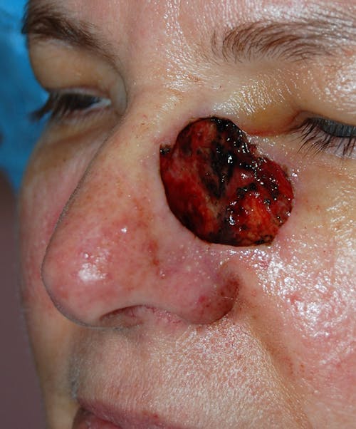
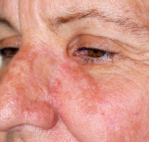
I’m amazed and grateful to say that what could have been a serious disfigurement is now, four months later, hardly noticeable
“I had a fairly large area of skin cancer removed from the side of my nose—about the size of a quarter. Dr. Gupta did a great job putting everything back together. I’m amazed and grateful to say that what could have been a serious disfigurement is now, four months later, hardly noticeable. Dr. Gupta is extremely skilled, and also personable and professional. Thank you, Dr. Gupta!”
5 star rating
Dr. Gupta performed the reconstruction, and I not only have no cancer, but I have my smile back!
“Dr. Gupta and his team are amazing. I had a serious squamous cell cancer treated with Mohs surgery, and I lost over half of my upper lip. Dr. Gupta performed the reconstruction, and I not only have no cancer, but I have my smile back!”
5 star rating
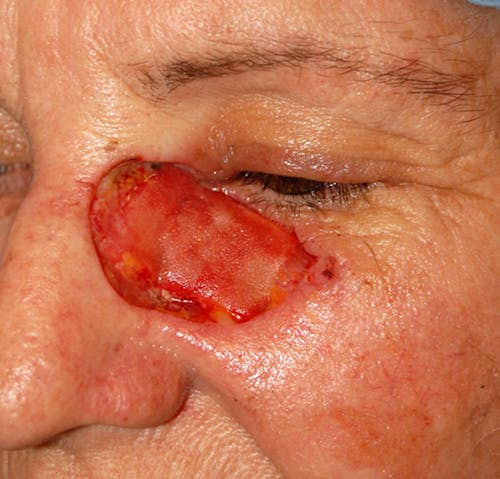
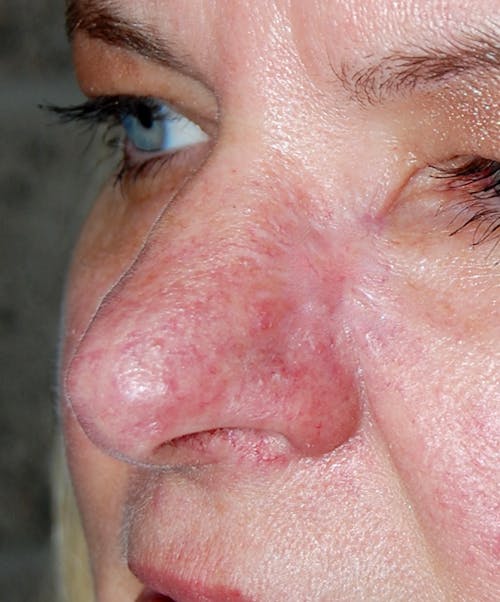
I was referred to Dr. Gupta by my dermatologist to repair my nose following Mohs surgery for skin cancer. Dr. Gupta rebuilt my nose, and I’m very pleased and relieved with the result
“Dr. Gupta rebuilt my nose, and I’m very pleased and relieved with the result. He was kind, highly capable, and instilled confidence throughout the entire process. From the initial consultation through surgery and post-op checkups, he was always prompt, courteous, and—most importantly—highly skilled and professional. I would highly recommend him.”
5 star rating
People were making comments like: 'You can’t even tell,' and 'He did an amazing job.'
“Dr. Gupta removed cancerous basal cells on my face; the procedure required ten stitches, and after two weeks, people were making comments like: 'You can’t even tell,' and 'He did an amazing job.”
5 star rating
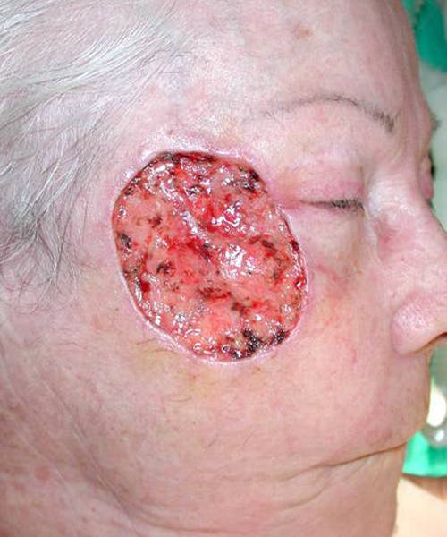
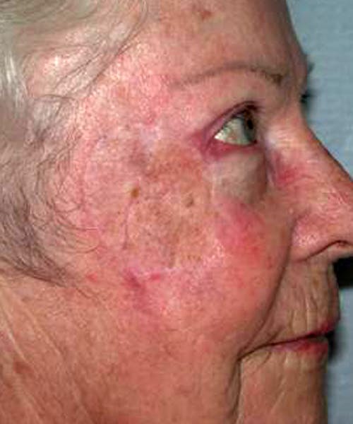
I am very happy with the results!
“I had to patch up a hole in my head left from cancer surgery, and I am very happy with the results!”
5 star rating
Results are better than expected
“The entire process with Dr Gupta and his staff has been excellent. Results are better than expected”
5 star rating
Excellent care. Warm and friendly people
5 star rating
He is very knowledgeable and friendly
“He is very knowledgeable and friendly, answering all our questions and concerns. It was a great experience.”
5 star rating
Dr. Gupta did a great job for both of us with no issues
“My wife and I both had some work on our faces. Dr. Gupta did a great job for both of us with no issues.”
5 star rating
Dr Gupta addressed my stretched earlobe painlessly and skillfully
“Great service and results! Dr Gupta addressed my stretched earlobe painlessly and skillfully.”
5 star rating
Dr. Gupta, thank you for your special attention to me!
“Briana, Kayla, and of course, Dr. Gupta, thank you for your special attention to me! Always positive with wonderful smiles and attentive care.”
5 star rating
Dr Gupta and his team are amazing!
“Dr Gupta and his team are amazing! They are all very supportive and helpful.”
5 star rating
Everyone in the office is very friendly and helpful
“Everyone in the office is very friendly and helpful. Dr. Gupta is very kind and explained everything very thoroughly.”
5 star rating
Dr. Gupta is professional, kind, and I trust him with my treatment completely
“Dr. Gupta is professional, kind, and I trust him with my treatment completely. He is very informative and always answers my questions.”
5 star rating
I highly recommend him
“Dr. Gupta and his staff are always patient and answer all the questions you may have. I highly recommend him.”
5 star rating
He listened with no judgment
“He listened with no judgment and was interested not only in my concerns, but ME! I was immediately put at ease by his team.”
5 star rating
Dr. Gupta provided me with a treatment plan within my budget
“Dr. Gupta provided me with a treatment plan within my budget. His office staff is amazing; it's truly like being with family.”
5 star rating
Dr. Gupta makes me look like a model
“I have been doing jazzercise for four years after gastric bypass; I lost all my weight but not the sag. Dr. Gupta makes me look like a model.”
5 star rating
He assured me that we would have great results, and I look fabulous!
“I lost over 100 lbs. the old-fashioned way, and Dr. Gupta was wonderful. He assured me that we would have great results, and I look fabulous!”
5 star rating
I had one surgery a year ago and had outstanding results
“After a large weight loss I decided to have excess skin removal surgery. I had one surgery a year ago and had outstanding results.”
5 star rating
I could not be happier with the result
“I was referred for abdominal reconstructive surgery as a result of multiple previous surgeries. I could not be happier with the result.”
5 star rating
I am so far happy with the results. I cannot recommend him enough!
5 star rating
He did amazing work with his precise skills!
“So glad I went with Dr. Abhay Gupta for scar revision. He did amazing work with his precise skills!”
5 star rating
My revised scar is flat and neatly done. I wish I had met him earlier.
5 star rating
He did a wonderful job on my surgery
“He did a wonderful job on my surgery. The staff here are professional and very quick in their responses.”
5 star rating
I flew into town from Seattle just for his services
“I flew into town from Seattle just for his services. I have been getting Botox for years, and he was just wonderful! I also upgraded this time around to lip fillers.”
5 star rating
Best Botox in San Diego!
“Best Botox in San Diego! Always pleased. The staff is amazing too!”
5 star rating
I had Botox by Dr. Gupta and he did an amazing job
“I had Botox by Dr. Gupta and he did an amazing job - much better than other places I have had it done before. Highly recommend!”
5 star rating
The best Botox experience I have ever had
“I went in to see Dr. Gupta for Botox and it was the best Botox experience I have ever had. I was so happy with the results and will definitely be back.”
5 star rating
No other Doc has made me feel so comfortable and welcome
“No other Doc has made me feel so comfortable and welcome. He is extremely professional, and the office is so relaxing and beautiful!”
5 star rating
I came in for Botox and filler and I couldn't be happier
“I came in for Botox and filler and I couldn't be happier. It is so natural and everything I was looking for. Thank you so much Dr. Gupta and staff!”
5 star rating
I trust him with my treatment completely!
“Dr. Gupta is professional, kind, and I trust him with my treatment completely! He is very informative.”
5 star rating
Dr. Gupta is professional, caring, and considerate
“Dr. Gupta is professional, caring, and considerate. Every physician in San Diego should take lessons from him.”
5 star rating
Always positive with wonderful smiles and attentive care
“Always positive with wonderful smiles and attentive care, I appreciate that you make me feel like I’m the only one in your care!”
5 star rating
Dr. Gupta is soft-spoken, calm, and quietly self-confident
“Dr. Gupta is soft-spoken, calm, and quietly self-confident. He answers all questions directly, which I appreciate.”
5 star rating
His staff is super, knowledgeable, and helpful
“He provides a lengthy print-out explaining what would be involved and what to expect. His staff is super, knowledgeable, and helpful.”
5 star rating
The laser resurfacing was wonderful on my face!!
“The laser resurfacing was wonderful on my face!! It helped take away the brown spots and sun damage I had!”
5 star rating
It gave me a fresh, tighter look!
“It gave me a fresh, tighter look! Recovery is pretty minor and the results were great!”
5 star rating
Dr. Gupta is so kind and helpful
“Dr. Gupta is so kind and helpful, always answering everything straightforward and in detail.”
5 star rating
I am always more than pleased with Dr. Gupta and his treatments on me
5 star rating
Skin Cancer Frequently Asked Questions
Will my scars be visible?
Dr. Gupta prioritizes discreet scarring in all of his procedures. During your reconstruction, he’ll place incisions along natural folds or hidden areas and use advanced closure techniques to minimize visible signs of surgery.
Can I have reconstruction later instead of immediately?
Yes. In some cases, Dr. Gupta may delay reconstruction until a final pathology report confirms that all cancer has been removed. Ultimately, the reconstruction strategy depends on safety and your goals.
Will my reconstructed skin feel different?
Yes, your sensation may be reduced or changed in the immediate area, especially if your nerve supply was disrupted.